3.2
Tissues of the teeth
This section describes the tissues present in teeth. They are enamel, dentin, cementum and dental pulp. These are common to all teeth, so we will look at them in more detail.
+
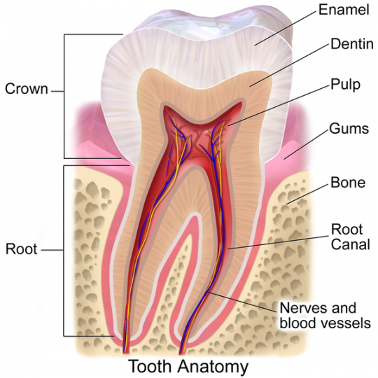
Source: Author BruceBlaus, Tooth Anatomy. See a full animation of this medical topic., license Creative Commons BY 3.0
Fig. 4. Tooth anatomy
3.2.1
Enamel
Enamel covers the anatomic crown of tooth. Enamel is translucent and appears in various colours – from yellowish to greyish white. The variations in colour are due to various factors, e.g. the quality of the crystal structure or enamel thickness.
It is the hardest tissue of the human body. Due to its hardness, along with the arrangement of oral tissues, teeth can sustain tremendous pressures of mastication. Enamel also protects the underlying dentin layer against thermal and chemical stresses. Enamel consists of about 96 % inorganic matter. The rest is made up of 1 % organic matter and 3 % water. All these percentages vary. The inorganic portion consists mainly of two principal minerals - calcium and phosphorus. They appear in the form of hydroxyapatite. There are also other elements, for example, fluoride and magnesium. The organic matter consists of proteins, carbohydrates and lipids.
Enamel structure is determined by enamel prisms, that look like hexagonal rods. There are millions of enamel prisms in enamel. Each prism starts at the dentinoenamel junction and extends to the surface of the tooth. However, the top layer is without prisms.
Enamel prisms are formed by ameloblasts. As these epithelial cells lose their regrowth ability after the completion of the crown, enamel cannot repair itself. The layer of enamel is up to 2 or 2,5 mm thick on the occlusal surface. It is thinnest at the neck.
It is also worth noting that enamel is permeable. This means that liquids can penetrate inside the enamel which acts as a semipermeable membrane. Thus, pigments contained in beverages can stain the enamel. More importantly, the condition of enamel itself can be altered chemically.
3.2.2
Dentin
Dentin makes up the largest portion of the tooth. Its thickness is up to 3 mm. Dentin, together with cementum encloses the pulp cavity. It, on the other hand, is surrounded by enamel in the crown area and by cementum in the root area. Its colour is light yellow and it is very porous. It can be permeated by liquids. It is harder than bone, but obviously not harder than enamel. The inorganic matter represents about 70 % of dentin and the organic about 30 %. Water is also present. The inorganic matter consists of hydroxyapatite crystals of calcium phosphate just as enamel. However, its structure is not prismatic. Another difference is that dentin is a living tissue. It is sensitive to various stimuli and can be repaired (see below for details).
Dentin is formed by cells called odontoblasts. Odontoblast processes run from the pulp cavity to the cementoenamel junction through dentinal tubules that give dentin its structure. They are more crowded near the pulp. The processes are responsible for dentin’s sensitivity to stimuli, such as pain, touch, thermal changes, and chemical substances.
Odontoblast processes are able to perform another important feat. They are able to form new dentin for as long as the nourishing pulp is healthy. As a matter of fact, as dentin grows, the pulp cavity becomes more narrow. In case of enamel damage, odontoblast processes can step in and form so called secondary dentin, a kind of reparative dentin. Unfortunately, it has its limitations and it is not often seen to completely seal over decayed enamel.
3.2.3
Cementum
Cementum is the part of the tooth that covers the roots. It covers dentin as a thin layer of tissue similar to bone in structure and composition. Just like dentin, it is yellow, but somewhat lighter. Cementum has 55 % organic material and water and 45 % of inorganic matter. The organic component includes collagen and proteoglycans. The inorganic matter consists mostly of calcium salts. Cementum joins enamel at the so called cementoenamel junction. It doesn’t mean that where cementum ends, enamel begins, and vice versa. The truth is, that in most cases, cementum is formed over the edge of enamel. However, in some teeth there is a gap between the two which makes the teeth sensitive. The neck of the tooth is also where cementum is very thin.
Cementum is the only part of the tooth which is also considered a part of periodontium. This is because the main function of cementum is to provide attachment points for the fibres of periodontal ligament which hold teeth in their place. This is not a one-time process. Cementum is formed continuously and new fibres attach to it. The main collagenous fibres that run from cementum to the alveolar bone are called Sharpey fibres. If need be, the fibres can be removed by cementum in response to tooth movements. This is the second function of cementum.
Cementum is formed by cementoblasts, but they are not present in some cementum due to the fact that there are actually several kinds of cementum, each in different layers. Only the cementum layer placed at the apex of the roots contains cementoblasts, along with collagen fibrils.
3.2.4
Dental pulp
The dental pulp is located in the pulp cavity (chamber). The cavity is both in the crown and in the roots. Hence, the pulp in the crown is called coronal pulp and the pulp in the roots is called radicular pulp. In the crown area, there are so called pulp horns under the cusps. The pulp grows smaller during life due to the expansion of dentin.
The pulp is a soft, jelly-like tissue. It is composed of 25 % organic matter and 75 % water. The pulp develops from the connective tissue and contains blood vessels, nerves and even lymph. These all enter the pulp cavity through apical foramina (singular foramen), openings at the apex of the roots. They are from 0.3 to 0.6 mm wide. Apart from apical foramina, there can also be other accessory canals, located laterally to the root region. Thanks to its composition dental pulp is often called the nerve of the tooth.
Different types of cells are present in the pulp. Fibroblasts are the most prevalent cells and are concentrated in the pulp mass. On the periphery, there are odontoblasts. Finally, there are also defensive and replacement cells of the connective tissues.
The functions of the dental pulp follow from the aforementioned information. The pulp nourishes odontoblasts and also replaces non-functional odontoblasts. Through these actions it helps form dentin. It also sends pain signals in cases of irritation by mechanical, chemical, or thermal agents. Since pulp is sensitive, dentists must be very careful when preparing crown stumps. For example, the heat generated by drilling can cause great irritation. Actually, the damage to the pulp may eventually prove irreversible and devitalize the tooth, although that may not become manifest for years.
Table 3. Vocabulary Table No. 3
English | Czech | English | Czech |
tissues | tkáně | enamel | sklovina |
dentin | zubovina (dentin) | cementum | cement |
pulp | dřeň | translucent | průsvitný |
arrangement | uspořádání | sustain | vydržet, snést |
layer | vrstva | prisms | prizmata |
rods | hranoly | junction | hranice, spojení |
permeable | propustný | beverages | nápoje |
stain | vytvořit skvrny | tubules | kanálky |
feat | počin | nourishing | vyživující |
reparative | opravný | seal over | uzavřít |
gap | mezera | fibres | vlákna |
jelly-like | rosolovité | cusps | hrbolky |
horns | rohy | apical foramina | hrotové otvory |
stump | pahýl |
©
For licensing reasons, this interactive object cannot be directly incorporated into the material. Click HERE to see the object.
Interactive object 5. Click on the part (dot) whose name appears above the picture (you will be directed to an external site).