2.2
Facial bones
Facial bones are not in touch with the brain. There are fourteen in total, out of which six are paired and two are single. The paired bones are maxillae, palatine, zygomatic, lacrimal and nasal bones and two inferior nasal conchae. The single bones are the vomer and the mandible. The mandible is the only moveable bone. The facial skeleton gives the face its basic shape. Its bones provide attachments for muscles that enable jaw movements and facial expressions. Facial bones also support the teeth. We start with a more detailed description of the maxillae and mandible, which are of great importance to the dental technician.
2.2.1
Maxillae
Unlike the lower jaw, the upper jaw is formed by two bones – the maxillae bones. However, they are joined in the midline of the jaw and they function as one bone. The maxillae are the largest bones of the face.
The maxilla consists of a body and four processes, zygomatic, frontal, alveolar and palatine. The body of the maxilla is made up of thin bony plates. The processes form a large portion of the hard palate, the floor of the nose, the walls of the orbits and alveolar tooth sockets. There are eight tooth sockets in each maxilla. Masticatory pressures are transferred by the processes to the cranial vault.
There are several openings within the maxilla and also the largest paranasal sinus, the maxillary sinus.
+
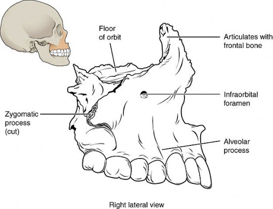
Source: Author OpenStax College, Illustration from Anatomy & Physiology, Connexions Web site., license Creative Commons BY 3.0
Fig. 2. Maxilla
©
For licensing reasons, this interactive object cannot be directly incorporated into the material. Click HERE to see the object.
Interactive object 2. Click on the part (dot) whose name appears above the picture (you will be directed to an external site).
2.2.2
Mandible
The mandible is a single bone which forms the arch of the lower jawbone. It is the longest and strongest bone. Also, thanks to the temporomandibular articulation, or joint (TMJ), it is the only movable bone of the head. There is a TMJ on both left and right side of the mandible. The movements of the lower jaw are performed by several muscles that extend from the skull to the mandible.
The mandible consists of the body and two rami (singular, ramus). The body is horseshoe-shaped. It forms the horizontal front and lateral sides. At the centre of the body, there is a mental protuberance, or chin. The body of the mandible can be divided into two parts – the base and the alveolar process. The latter part is where the socket of mandibular teeth are found. The upward motion of the mandible makes the maxillary and mandibular teeth meet (occlude).
The two rami are parts of the mandible that extend vertically (they are upward sloping) on both sides at the ends of the body of the mandible. They do so at a place called the angle of the mandible. Each ramus has two important upper ends, the condyloid and coronoid processes.
+
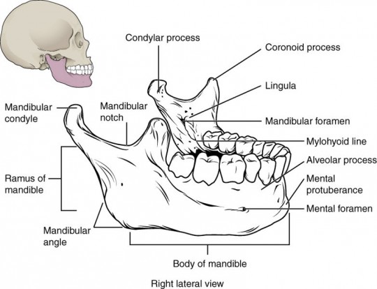
Source: Author OpenStax College, 726 Mandible, license Creative Commons BY 3.0
Fig. 3. Mandible
The condyloid process is the rear end of the ramus. It articulates with the mandibular (or glenoid) fossa of the temporal bones to form the TMJ. Its name is derived from Latin and means knuckle-shaped. The second process, the coronoid process, is placed in front of the condyloid process. It enables the attachment for the temporalis muscles whose role is to lift the mandible. The depression between the two processes is called the mandibular notch.
There are two sets of foramina in the mandible – mandibular and mental foramina. The mandibular foramina are located in the middle of each ramus on the inside, at the level or slightly above the level of occlusal surfaces of the teeth. Through these openings pass alveolar nerves and blood vessels supplying the roots of the mandibular teeth. In this area, the dentists can inject anaesthetic to desensitize (numb) the teeth in the corresponding side.
The mental foramina are not located directly on the chin. Instead, they are located on the lateral facial surfaces of the mandible, usually below the second premolars. These openings transmit the mental nerve and blood vessels, some of which supply the chin. The position of the foramina changes in edentulous people due to bone resorption.
©
For licensing reasons, this interactive object cannot be directly incorporated into the material. Click HERE to see the object.
Interactive object 3. Click on the part (dot) whose name appears above the picture (you will be directed to an external site).
2.2.3
Palatine bones
The palatine bones resemble the letter L in their shape. They can be found behind the maxillary bones, with which they are joined. They form the posterior part of the hard palate, the floor of the nose, and a portion of the orbits. The palatine bones contain one large foramen and several smaller foramina. All the openings allow nerves to pass through and the large foramen also gives space to descending vessels.
2.2.4
Zygomatic bones
The zygomatic bones are called cheek bones in layman’s terms. The temporal process of each zygomatic bone extends to the zygomatic process of each temporal bone. Thus a zygomatic arch is formed on each side of the skull. The processes are used to transfer masticatory forces from the maxillae to the cranial vault. Zygomatic bones also help to form the lateral sides of the orbits.
2.2.5
Lacrimal bones
The lacrimal bones, or the lacrimals, are small, thin and thus fragile bones that form the front section of the medial wall of each orbit. In each lacrimal bone, there is a groove that allows tears to flow into the nasal cavity.
2.2.6
Nasal bones
The nasal bones are positioned next to each other and together they form the bridge of the nose (nasal septum). These bones give the nose its shape.
2.2.7
Inferior nasal conchae
The inferior nasal conchae are fragile, curved, scroll-like bones that are placed below middle nasal conchae which are part of the ethmoid bone. They provide support to mucous membranes which adapt (warm, moisten, purify) the outside air for inhalation.
2.2.8
Vomer
The vomer bone is one of the two single bones in the facial area. It is a thin, flat, elongated bone. Together with the ethmoid bone, it supports the septal cartilage and thus forms the nasal septum.
Table 2. Vocabulary Table No. 2
English | Czech | English | Czech |
palatine | patrová | zygomatic | lícní |
lacrimal | slzní | inferior | spodní |
vomer | radličná | vault | klenba |
motion | pohyb | extend | vybíhat |
rami | ramena | angle | úhel |
condyloid process | kondylární výběžek | rear | zadní |
knuckle-shaped | ve tvaru kloubu (na prstu ruky) | coronoid process | koronoidní výběžek |
notch | incisura | edentulous | bezzubí |
resemble | připomínají | fragile | křehké |
groove | brázda | mucous membranes | sliznice |
cartilage | chrupavka |

Video 2. Facial bones
©
For licensing reasons, this interactive object cannot be directly incorporated into the material. Click HERE to see the object.
Interactive object 4. Click on the bone (dot) whose name appears above the picture (you will be directed to an external site).