1.1
Tools and equipment of a surgery
A dental office is equipped with a dental chair, which is a foot or manually operated. It consists of seat, backrest and an adjustable headrest. There are also unit with air and water syringes, saliva ejector, an abrasor, a cuspidor, a light, X-ray and a stool. We can find different kind of furniture, which is given close to a dental chair. These are table or cupboard with sink, sharpening stone, computer, and autoclave. Tray with instruments (an explorer, a probe, a scaler, cotton pliers, tweezers) is usually on the piece of cupboard. Another cupboard is for instrument and material storage.
+
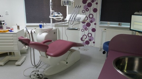
Fig. 1. A dental hygienist surgery with dental chair
Each instrument must be recognized by sight and distinguished at a glance by the profile of the instrument on the sterile tray. The dental hygienist must know the names and associate each tool with various phases of instrumentation.
We classify the instrument according its purpose and use to examinational instruments – probe, explorer and treatment instruments – curettes, scalers (a sickle, a hoe, a chisel).
1.1.1
Instrument parts
There are three major parts, the working end, the shank, and the handle.
- Working end – it is a part which carry out the purpose and function of the instrument. Each working end is unique to the particular instrument. They can be either sharp, then this working end is called a blade, or non-sharp, then it is called a dull blade.
- Shank – it connects the working end with the handle. They can have different shapes and rigidity according to their purpose. Straight shank is used to tooth surfaces for anterior teeth. Angled shank is to tooth surfaces with restricted access, such as proximal surfaces of posterior teeth. Rigid shank is designed for removal of heavy calculus deposits. It is thick to bear pressure without flexing. Less rigid shank, more flexible, is used for removal of fine deposits of calculus.
- Handle – it is the part which is grasped during activity. It can be single end instrument, double-ended instrument (mirror image), or cone socket handle, which is separable from the shank and allows exchange of shank with working end.
1.1.2
Examinational instruments
Parts of the gingival and dental examination are made by direct visual observation, while other parts require tactile examination using a probe and an explorer. They are assisted by a mouth mirror.
Probe
A probe is smooth, slender instrument usually round in diameter with a rounded tip used to make the initial assessment. Dental hygienist measures pocket depths around a tooth. They insert the tip of probe into the gingival sulcus, down to the base of the pocket. The first marking above the pockets indicates its depth. The markings are calibrated in millimetre increments.
+

Fig. 2. Probe WHO
A probe is used to:
- determine the periodontal status, classification of diseases such as gingivitis, periodontitis, or determination of the extent of inflammation.
- make a pocket survey. It examines the shape, dimension and measures visible gingival recession.
- evaluate gingival bleeding together with bleeding index.
- create a treatment plan from all previous points mentioned above.
- evaluate the condition after treatment and self-treatment.
Furcation probe is used for probing the extent and depth of furcation lesions from different angles.
Explorer
An explorer is a slender stainless-steel instrument with a sharp point used for examination of the surfaces of the teeth to detect irregularities. Dental hygienist distinguishes tactile sensations when exploring.
They can feel:
- tooth structure – normal anatomic configurations
- restored surfaces – smooth surfaces (amalgam, plastic, gold)
- deposits – calculus
- restorations – over contoured, irregular margins, rough surface
- demineralized or carious lesions, abrasion, erosion
Dental hygienist must work very gently because penetration can cause cavitation in places which could be remineralized.
Mouth mirror
It visualizes the certain area of the mouth where visibility is difficult or impossible. The mirror surface can be flat, it produces a double image, or concave, it is magnifying, and front surface, which eliminates “ghost” images. Disadvantage is that it tends to become stretched.
It usually has thicker handle to a more comfortable grasp.
+
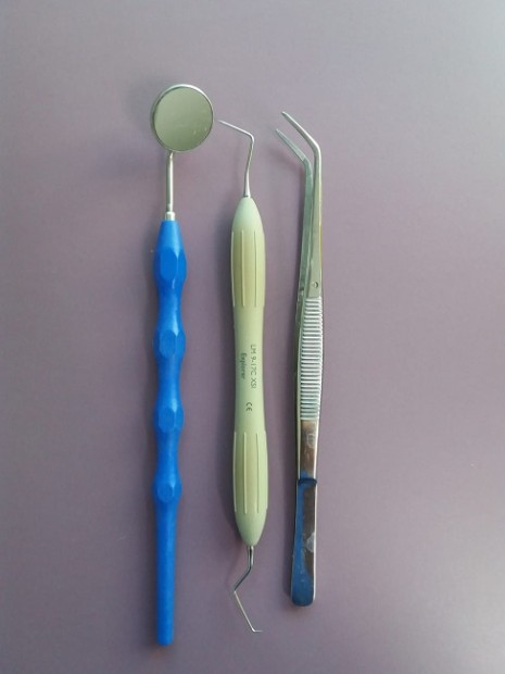
Fig. 3. Basic examination set
1.1.3
Treatment instruments
Each instrument is designed for a specific type of application during treatment procedures. Scalers are designed for supragingival treatment procedures and curettes are then for subgingival treatment.
Curette
A universal curette can be adapted for instrumentation on any tooth surface. Common purposes and uses are:
- sublingual scaling and root planning
- finishing of procedure after ultrasonic or sonic scaling
- removal of supragingival calculus
- curettage of the inflamed lining of the gingival wall of a pocket
- obtaining a sample of subgingival plaque
Gracey curettes
They are designed specifically to be site specific to remove supra and sublingual calculus. Blades are paired on the sides of the instrument, and their design allows better adaptation to the anatomy of the roots. They are identified by a number in the handle. Each instrument has an odd number to identify each blade.
+
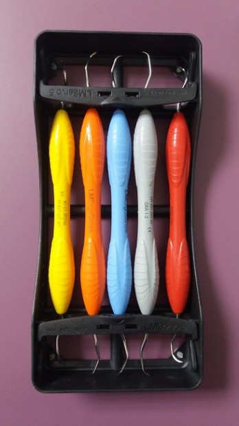
Fig. 4. Gracey currete
Scalers
Scaler is an instrument used to remove calculus from the teeth. It has scraping edges and sharp tips to access tight spaces between teeth. The working ends are found in various shapes and sizes.
Sickle scaler (curved scaler), two cutting edges on a curved blade, is used mainly to remove supragingival calculus and calculus slightly below the gingival margin.
Hoe scaler, single, straight cutting edge, is used for removal of gingival calculus, especially large, accessible, tenacious pieces. It is also suitable for deep, narrow pockets and concave root surfaces, for both vertical and horizontal techniques.
+
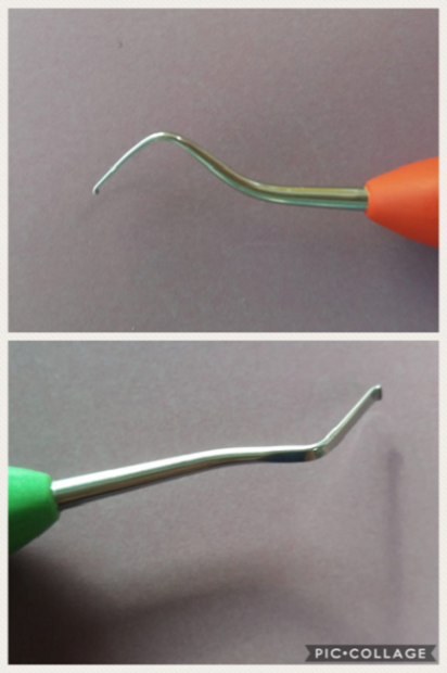
Fig. 5. Hoe scaler anterior (green), posterior (orange)
Chisel scaler with single straight cutting edge is good for removing supragingival calculus from exposed proximal surfaces of anterior teeth where interdental gingiva is missing. When the lips and cheeks are flexible and permit retraction for proper position of cutting-edge scaler then it can be used for proximal surfaces of premolars.
File scaler has multiple cutting edges lined up as a series of miniature hoes on a round, oval, or rectangular base. It is considered as a supplementary instrument and usually used during scaling and root planning.
Power scaler provides effective treatment, the tip moves in a back-and-forth motion. It is a kind of ultrasonic instrumentation. Dental hygienists move tip along the side of a tooth and direct the vibration right onto the calculus without knocking the enamel or cementum.
Air scaler is powered by the air.
©
For licensing reasons, this interactive object cannot be directly incorporated into the material. Click HERE to see the object.
Write the correct translation (the user will be directed to an external page)
1.1.4
Instrumentation technology
Summary
Imaging techniques are used to detect caries in their initial state, showing the roots of the tooth, position of teeth, etc. Sterilization and disinfection are very important in infection control. Manual cleaning of instrumentation is difficult and time-consuming procedure.
X-ray
About one third of the tooth decay is found by a dentist or dental hygienist during a regular examination by a probe and dental mirror. Very small defects that do not cause difficulties can be discovered by regular X-ray examination. It represents another third of discovered tooth decay.
Most often X - ray is OPG (OrthoPantomoGram) frame that is made by extraoral imaging technique. It is the only imaging method, which is able to depict both jaws in the picture with the associated cavities, teeth, and both jaw joints.
+
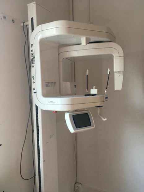
Fig. 6. OPG
BW (BiteWing) images is made by intraoral technique. BW picture captures the crowns of the teeth of both jaws with the appropriate alveolar bone.
+
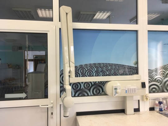
Fig. 7. Intraoral X ray
The modern RTG devices are equipped with a flat-panel detector that converts the incident radiation onto a computer signal and the X-ray image of the subject immediately appears on the monitor.
Advantage
This image has certain advantages include not only the ease of archiving the images but also the distribution of images, then possibility of subsequent editing (adjusting brightness, magnification, distance measurement, angles measurement) which leads for better evaluation by an expert.
+
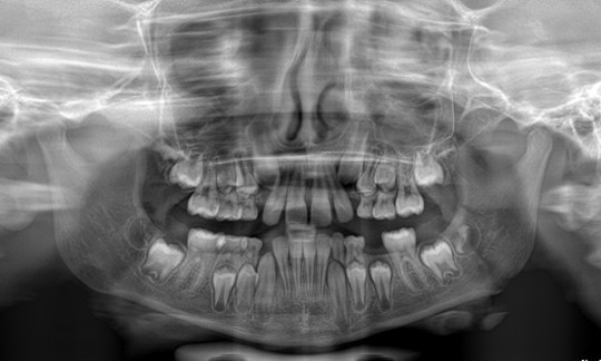
Fig. 8. X ray picture
Disadvantage
Absorbed X-rays have negative effects on the body. These can range from acute illness from irradiation or local effects on the skin to genetic cell transformation.
X-rays can only be used based on a medical indication, and the patient should not be exposed to radiation unreasonably.
Intraoral camera
Intraoral camera is the optical imaging system. It transmits multiple-magnified image of the oral cavity directly on the LCD screen, located on a dental chair. This miniature camera allows dental hygienist to look at the places hardly accessible by sight. Dental hygienist using a high-quality intraoral camera can examine the patient, the caries is displayed through camera like a dark shadow. The device can consist of camera, a light guide and a tip. Camera is connected via a USB connector with a monitor that is capable of storing record examination. The camera makes a snapshot that can be saved in documentation. Everything is mediated by the software that is supplied with the device.
Diagnodent pen
The intraoral scanner uses laser fluorescence method which allows detection of caries in the period of their development. The success rate of the finding of caries is very high. It is a diagnostic device based on the infrared laser principle fluorescence. Firstly, the teeth must be cleaned. The examination is painless and lasts 5 – 8 minutes. Dental hygienists move with probe over the tooth surface in all directions so that they detect the maximum value of the tooth. Measured values for each tooth are recorded in the documentation. Value 0 -13 means healthy teeth, 14 – 20 the beginning of caries, 21 – 29 is caries in enamel, 30 and more means caries in dentine.
Digital photo
The digital camera records the image on digital chip. The photos are applied in patient documentation before, during and after treatment.
All items and surfaces must be cleaned easily to prevent cross-infections. Dental hygienist must protect himself and her/ his clients. Dental hygienist must wear uniform, protective gloves, and mask. She should have short, unpolished nails and hair fastened back. Client should postpone their appointment in case of illnesses and should have clean teeth.
The equipment of surgery is either disposable (needles, plastic cups, chair coverings), it can be used once and then is discarded, or reusable, it can be used again, but definitely it must be washed (uniform, towels), disinfected (hand pieces of unit, mirrors), or autoclaved (mirrors, scalers, probes).
Autoclave
Sterilization is the process by which all forms of life are destroyed. It can be done with moist heat, dry heat, or chemical vapour sterilizer. Autoclave is a pressure chamber which is used in medical applications to perform sterilization. The equipment is pressurized by saturated stem at high temperature for 20 minutes.
+
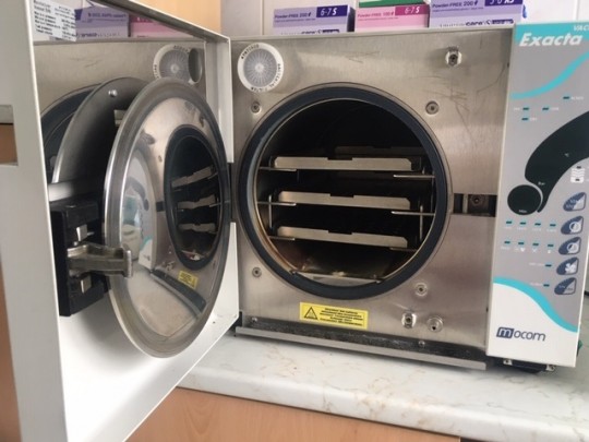
Fig. 9. Autoclave

Instrument cleaning instruction (the user will be directed to an external page)
©
For licensing reasons, this interactive object cannot be directly incorporated into the material. Click HERE to see the object.
Match the correct words (the user will be directed to an external page)
Vocabulary
English | Czech | English | Czech |
at a glance | na pohled | tray | tácek |
instrument | nástroj | tool | nástroj |
explorer | pátradlo | dental mirror | zubní zrcátko |
scaler | skaler, škrabka | probe | sonda |
capture | zachytit | cotton pliers | zubní pinzeta |
saliva ejector | odsávačka | cuspidor | plivátko |
adjustable | polohovatelný | stool | stolička |
air and water syringe | vzduchová a vodní pistole | waste container | odpadkový koš |
access | přístup | slender | štíhlý |
pocket | kapsa | depth | hloubka |
insert | zavést | increment | přírůstek |
extent | rozsah | tactile sensation | hmatový pocit |
explore | prozkoumat | tenacious | soudržný |
narrow | úzký | concave | vydutý |
retraction | odtažení | hoe | motyčka |
subsequent | následující | adjust | upravit |
magnification | zvětšení | irradiation | ozáření |
value | hodnota | chamber | komora |
blade | čepel | curette | kyreta |